Service Overview:
Skeletal imaging is used to assess bones and joints, helping diagnose injuries, degenerative conditions, and structural abnormalities. This imaging method provides high-resolution X-ray scans that allow medical professionals to evaluate fractures, joint integrity, and bone health.
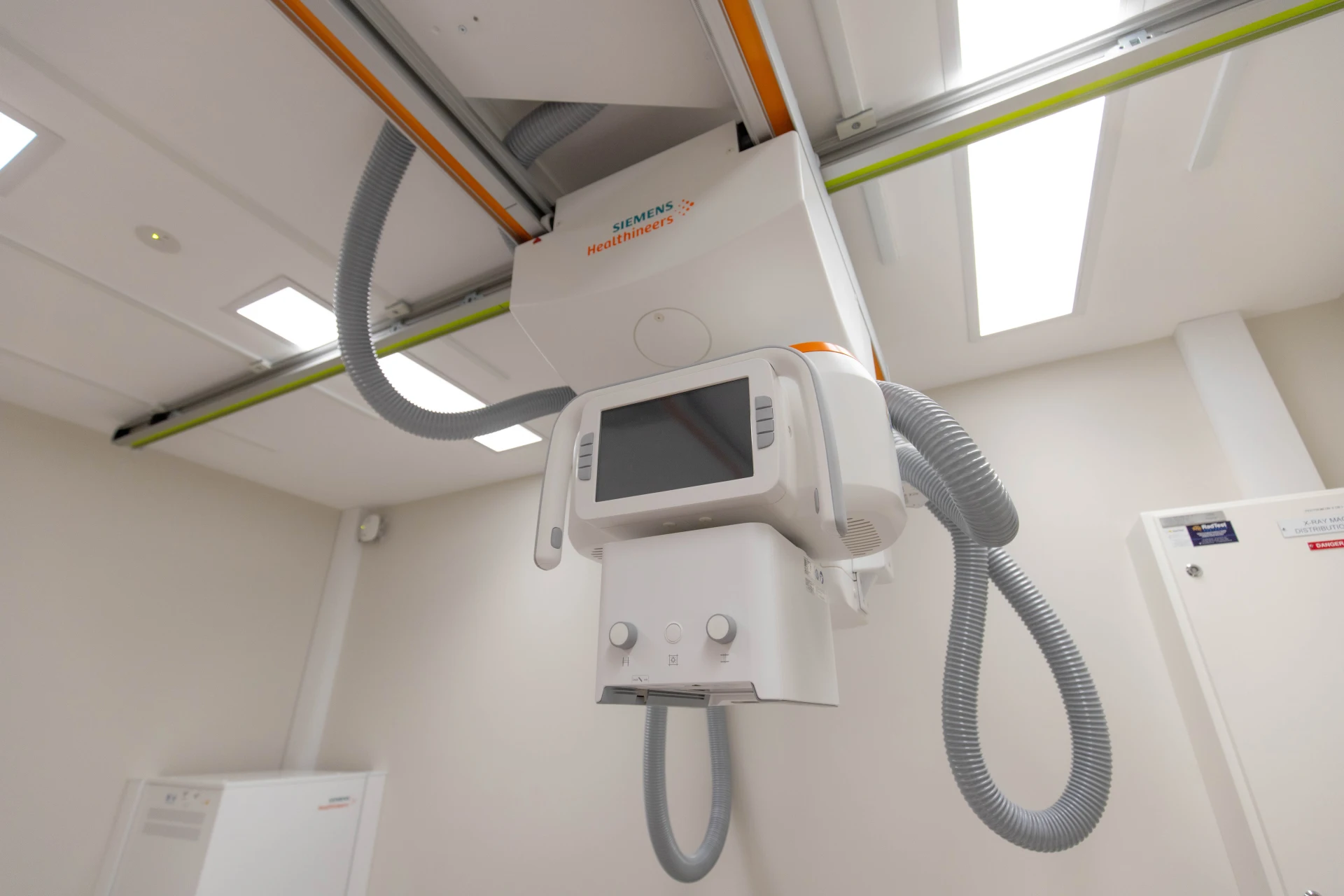
At Victorian Imaging Specialists, we use advanced digital imaging technology to capture clear and detailed skeletal images, ensuring accurate diagnosis while using minimal radiation exposure. Whether investigating an injury or monitoring a long-term condition, skeletal imaging is a critical tool in musculoskeletal healthcare.
Common Uses of Skeletal Imaging:
- Identifying fractures, stress fractures, and joint dislocations
- Assessing bone density and detecting early signs of osteoporosis
- Diagnosing arthritis, osteoarthritis, and joint degeneration
- Evaluating skeletal deformities and congenital bone conditions
- Monitoring recovery from bone injuries and post-surgical healing
What to Expect During the Procedure:
During the scan, patients may need to stand, sit, or lie down, depending on the area being imaged. The radiographer will carefully position the affected limb or joint to ensure the best possible images. Patients must remain still for a few seconds while the scan is taken. The procedure is completely non-invasive and typically takes only a few minutes to complete.
Preparation Guidelines:
For most skeletal imaging, no special preparation is required. Patients may be asked to remove jewellery, metal accessories, or clothing with metal fastenings, as these can interfere with imaging. A gown may be provided if necessary. It is essential to inform staff if there is any possibility of pregnancy so that appropriate precautions can be taken.
Safety Information:
X-ray-based skeletal imaging uses a very low level of radiation, and every step is taken to ensure patient safety. The benefits of early detection and accurate diagnosis far outweigh any minimal risks associated with radiation exposure.
Results and Next Steps:
Once the scan is completed, a specialist radiologist will review the images and provide a detailed report to the referring doctor. Patients should schedule a follow-up consultation with their doctor to discuss the findings and any necessary treatment options.
For more information or to book a skeletal imaging scan, please contact your nearest VIS clinic.
