
Overview
X-rays are a diagnostic tool that uses minimal doses of radiation to produce images of the body’s internal structures. They are particularly effective for examining bones, joints, and certain soft tissues, assisting in the detection of fractures, infections, and various medical conditions.
Common Applications
- Skeletal Imaging: Assessing bones for fractures, dislocations, or diseases.
- Chest X-rays: Evaluating the lungs, heart, and chest wall for conditions like pneumonia or heart failure.
- Abdominal X-rays: Identifying issues in the gastrointestinal tract, such as obstructions or swallowed objects.
Procedure
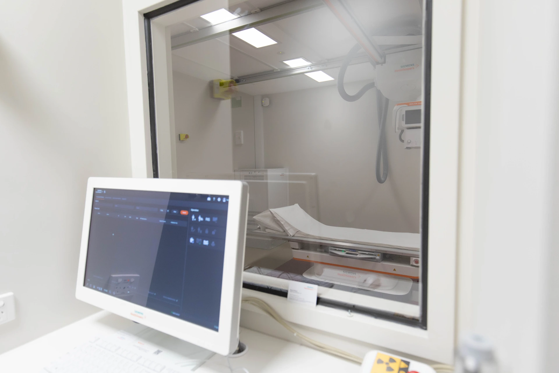
During the X-ray procedure, patients may be asked to stand, sit, or lie down, depending on the area being examined. Our radiographer will position the patient and the X-ray machine appropriately to capture clear images. It’s essential to remain still during the exposure to ensure image clarity. The process is quick, typically lasting only a few minutes.
Preparation
Generally, no special preparation is required for standard X-rays. Patients may need to remove jewelry or wear a gown if clothing could interfere with the imaging. It’s crucial to inform your radiographer if there’s any possibility of pregnancy, as precautions may be necessary.
Safety
While X-rays involve exposure to low levels of radiation, they are considered safe for most patients. The benefits of accurate diagnosis and treatment planning usually outweigh the minimal risks associated with radiation exposure.
Results
After the X-ray, a radiologist will analyse the images and provide a report to the referring physician. This process ensures that patients receive accurate diagnoses and appropriate care plans.
For more information or to schedule an appointment, please contact your nearest VIS clinic.